
IACC Strategic Plan
For Autism Spectrum Disorder
2016-2017

Introduction
Aspirational Goal: Discover how alterations in brain development and the function of physiological systems lead to ASD in order to enable the development of effective, targeted interventions and societal accommodations that improve quality of life for people on the autism spectrum.
Current scientific evidence suggests that ASD results from subtle alterations during brain development that affect brain structure, function, and connectivity. However, our knowledge about its causes remains incomplete and significant gaps in science have hindered attempts to develop therapies to improve quality of life for individuals with ASD. Over the course of the last decades, several studies have revealed the role of prenatal or perinatal stressors and genetic contributors to the risk of developing ASD, possibly acting through changes in early brain development. The biological mechanisms by which known gene mutations cause syndromic ASD (i.e., the subtypes of ASD that are usually caused by a single genetic abnormality) by altering the underlying neural circuitry of the brain are under intense study. These genetic variants are associated with remodeling of genetic material, changes to ion channels (which are the basis for cellular function and communication), and proteins that regulate cell-to-cell communication. Taken together, this research suggests there may be shared features of the underlying biology across the spectrum of autism. However, we currently know very little about the precise pathways that cause the circuit changes driving the core behavioral features in ASD, but new tools promise to accelerate this area of investigation. In addition, while there have been recent gains in understanding ASD developmental trajectories and the nature and prevalence of co-occurring conditions in persons with ASD, more work is needed to understand these aspects of ASD and develop strategies to target them successfully.
Molecular Mechanisms Affected by Genes Implicated in ASD
Genetic studies of ASD have identified more than 100 high-risk genes and estimate that several hundred additional genes of this type will be identified in the future.1 It seems likely that over 1,000 genes conferring lowerdegrees of autism susceptibility will also be identified in the future.2,3,4,3,6,7 At present, the known functions of these genes converge on biological processes important for neuronal communication and regulation of the expression of genes and proteins.
The discoveries of gene mutations that cause syndromic ASD (e.g., tuberous sclerosis complex (TSC), Rett syndrome, Fragile X syndrome, Phelan McDermid syndrome), and the dozens of rare de novo (spontaneous) mutations that disrupt gene function in ASD8 have enabled scientists to explore the biological effects of the various involved genes in cellular and animal experiments. This has led to an explosion of research examining how these mutations alter the biology of cells and investigating their effects on neural circuitry and behavior. On the horizon are genetic tools that will enable the introduction of mutations into non-human primates,9,10 which possess a much greater behavioral repertoire and a more human-like brain than rodents or other animal models. However, these experiments will need to be designed carefully because the numbers of matched control and mutant animals need to be very small as compared to rodent studies.
A major advance over the last few years is the ability to take skin or blood cells from persons with ASD, create induced pluripotent stem cells (iPSCs), and differentiate these cells into neurons, which can enable the study of neural function at the cellular level. This new technology allows scientists to study the effects of ASD mutations in human brain cells in addition to commonly used transgenic animal models. Furthermore, iPSCs are attractive models for identifying molecular phenotypes linked to syndromic ASD, but a more high-throughput means of identifying and validating their relevance to ASD is needed. A strategy for identifying relevant molecular phenotypes in iPSCs from the much more common idiopathic ASD (ASD of unknown cause) remains a daunting task. In addition, new research may make it possible to grow "brain organoids," which are clumps of brain tissue partially organized to have some features of the human brain, from iPSCs. These partially matured "mini-brains" can be grown in a culture dish and can be used to enable the study of the early development of brain structures that occurs in utero, as well as the cellular and circuit abnormalities related to ASD-linked mutations.11,12 However, these in vitro studies will introduce a number of variables related to culture conditions, and deliberate actions will be required to evaluate reproducibility.
Though new iPSC and brain organoid technologies allow for the study of human cells and circuits derived from persons with ASD, there is no substitute for careful structural and transcriptomic studies in postmortem tissue which remains exceptionally rare. Efforts need to be redoubled to increase the accessibility of brain tissue from well-characterized ASD cases. The establishment of collaborations like the National Institutes of Health (NIH) NeuroBioBank and Autism BrainNet facilitates the distribution of high-quality, well-characterized human postmortem brain tissue for the research community.
Enhancing efforts to increase public awareness about the value of tissue donation for understanding brain disorders like autism will most effectively advance the science.
Studies of postmortem brain tissue from persons with ASD have demonstrated decreased expression of sets of genes related to synaptic function, including many of the known ASD risk genes. Surprisingly, despite the 4:1 male to female ratio in ASD, these changes in synaptic gene expression were not more evident in males than females. However, there was an observed upregulation of genes related to microglial and astrocytic function (brain cells that provide support for neurons) that was more pronounced in males.13 These gene expression differences may help explain why ASD occurs more frequently in males. Moving forward, new gene expression mapping technologies have the potential to better characterize altered patterns of gene expression in specific brain cell types, offering the opportunity to precisely associate gene expression differences at a cellular level. In these studies, it will be critical to include an examination of gender-related differences in gene expression.
The impact of sex chromosomes on differences in gene expression between males and females – and how this may contribute to ASD – is also an area of research needing additional attention. The role of genes on the Y (male) and X (female) chromosomes extends beyond reproduction-related functions. Studies have suggested that genes on the sex chromosomes may act as broad regulators of gene expression. Therefore, differences in gene regulation by X-X gene pairs in females versus X-Y gene pairs in males may have different effects on the dosage-dependent expression of other genes, including those implicated in ASD-relevant molecular pathways.14,15,16
Another remaining challenge is to understand how the effects of hundreds of implicated genes converge to cause ASD’s common features. And conversely, more work is needed to determine how individual genes and their interactions with early life events explain the biological basis of the heterogeneity of ASD symptoms, which range from severe intellectual disability and absence of verbal language, to mild social deficits with normal cognitive function. With regard to the 4:1 male to female ratio in ASD mentioned above, further work is also needed to understand the phenotypic differences between girls and boys with ASD, and how these differences should inform development of screening and diagnostic tools, interventions, and services that meet the needs of both girls and boys on the autism spectrum.
Structure and Function of Brain Circuits in ASD
Structure and Function
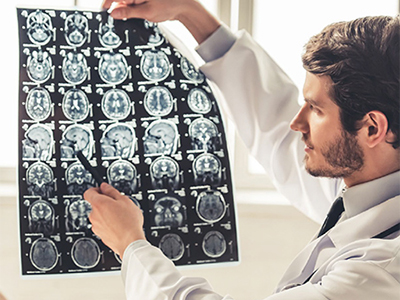
Autism is characterized by atypical patterns in physical brain connections (structure) and in how regions communicate with each other (function). Brain structure in individuals with ASD can be compared to typically developing children using advanced magnetic resonance imaging (MRI) techniques to measure size and shape of brain regions over time, as well as diffusion tensor imaging (DTI) to examine the structures of the major connections between brain regions. Brain circuit function can be investigated using non-invasive markers, such as functional magnetic resonance imaging (fMRI), magnetoencephalography (MEG), electroencephalography (EEG), and functional near-infrared spectroscopy (fNIRS). These studies have shown differences in activation patterns in individuals with ASD in response to sensory processing of visual, tactile, auditory, and verbal stimuli.17,18,19,20
Although non-invasive measures of brain connectivity have demonstrated differences between brain regions in persons with ASD compared to controls, it is unclear how these differences account for the heterogeneity in ASD symptoms. Earlier findings from fMRI, DTI, and pathological studies highlighted a pattern of reduced long-distance connectivity and increased local connectivity.21 While these principles still largely hold, newer research has revealed greater nuance and specificity.22,23
On the gross anatomical level, the brains of autistic individuals appear normal. Microscopic studies have reported disordered cell organization with smaller, more densely packed neurons in many regions of the limbic system, a part of the brain which is known to play an important role in learning, memory, and emotions. One of the more reproducible findings is a reduction in the number of Purkinje cells in the cerebellum.
Current evidence suggests that early brain overgrowth is present in approximately 15% of 2-year-old boys with ASD, but is much less common in girls.24,25 Brain enlargement relative to body size in this subset of boys persists at least through 5 years of age26 and is associated with lower language ability at age 3 and reduced intellectual ability at age 5.27 Currently, the prevailing theory is that early brain overgrowth normalizes during adolescence and adulthood,28 although this is based on cross-sectional studies. A truly longitudinal analysis, where the same individuals are imaged throughout life, is needed to clearly establish this. Studies at the cellular level have begun to describe differences in neuronal growth and organization in ASD brains.
Interestingly, recent studies have identified significant inter- and intra-individual variability in neural functioning in ASD. Heterogeneity in the ASD phenotype also contributes to greater functional variability within ASD groups. To address this heterogeneity, a greater number of studies are examining dimensional traits in large samples of ASD and neurotypical groups.
Ongoing work is linking these functional and structural differences to core features of ASD including studies on social communication, language, and restricted and repetitive behaviors. Recent work has identified neurobiological correlates of sensory processing in autism,29,30,31 including findings of reduced modulation of connectivity between the thalamus (a brain structure responsible for relaying sensory and motor signals to the cerebral cortex as well as regulating consciousness, sleep, and alertness) and cortical regions in response to sound or touch. The extent of this reduced connectivity was related to parent reports of sensory over-reactivity.30 In the domain of touch, reduced response was seen in ASD children in social-emotional brain regions to a soft caress compared to typically developing children, while increased activation was seen in response to non-caress-like touch in the brain’s primary sensory cortex. This over-activation may be related to the hyper-sensitivity to touch seen in some ASD individuals.
There is a need for a greater understanding of the relationship between intrinsic functional brain organization during rest and functional connectivity dynamics during task states. Additionally, there is a need for a better understanding of brain function and connectivity during tasks that better capture the complexity of real-world interactions for individuals with ASD.32,33
Circuit Activity in ASD
As is true of almost all brain disorders, the symptoms experienced by individuals with ASD are linked to alterations in brain circuit function. Alterations in the formation of brain circuits that occur in utero, during infancy, and in childhood can have long-lasting impacts on circuit function in adulthood. During these early critical time periods, connections between brain regions are dramatically molded by brain activity and may be altered by injury or inflammation.
Identification of genes involved in syndromic autism enable the study of the human phenotypes associated with those genetic syndromes as well as phenotypes of genetic animal models carrying the same mutations. The study of several forms of syndromic ASD have revealed nervous system differences such as differences in abundance of certain cell types, in neural circuits, and in brain activity.34 Environmental insults, for example those due to infection in utero, premature delivery, or perinatal cerebellar hemorrhage also alter the construction of brain circuits leading to ASD.
There are continuing challenges in applying animal models to understand the biology of autism. Because, in many cases, autism impacts uniquely human aspects of social-communicative behavior (e.g., spoken language), developing and measuring analogous phenotypes in animals has proven difficult. Because autism impacts brain regions not developed in some animal species, some neural circuitry is not readily amenable to study in these models. Moving forward, increased use of species more comparable to humans in both biology and behavior will be necessary. Notably, genetic tools previously limited in application to mice can now be applied in rats and even non-human primates, such as macaques and marmosets. Furthermore, the circuit alterations that have been described in ASD models vary considerably. Variability due to methodological differences among labs may diminish the value of the research findings. Incorporation of a more standardized and systematized approach to studying the altered circuits in ASD models could be valuable to the field.
Fortunately, new powerful technologies for interrogating and modulating brain circuits are revolutionizing neuroscience. The BRAIN Initiative is a multi-Federal agency, public-private partnership in the US to advance brain circuit neurotechnologies and also engage multiple international efforts. These technologies promise to expand the ability to understand brain circuit differences due to genetic and environmental influences that contribute to many diseases, including autism. The brain circuit alterations implicated in ASD in animal models can now be explored in detail using these new technologies to map neural connections over large expanses of brain,35 record from a large number of neurons during a behavioral task, and turn on or off specific types of neurons to understand the nature of brain circuit alterations caused by biological mechanisms tied to ASD.
Role of Immune System in Brain Development and ASD
Increasing evidence suggests immune dysregulation and neuroinflammation may be implicated in the severity and pathogenesis of the autism phenotype.36,37,38 One recent meta-analysis of 17 studies identified significantly altered concentrations of immune regulators known as cytokines in ASD patients compared to healthy controls, adding to the evidence of increased inflammatory signals in ASD.39 Despite many studies demonstrating altered levels of immune biomarkers and abnormal immune function in both the peripheral and central nervous system in ASD, it is not clear whether the immune system plays a direct role in the development of the disorder via an impairment of neurodevelopmental processes. Several recent studies suggest that maternal immunological factors may play a role in the pathogenesis of ASD during prenatal development.40,41,42,43,44,45,46
Microglia are innate immune cells that reside in the central nervous system and are activated in response to infection or inflammation. Even in their so-called resting state, they perform critical functions, including regulating the number of neural precursor cells,47 maintaining synaptic organization, and synaptic pruning (removing excess or underutilized synapses during development).48,49 Analyses of autism brain tissue reveal alterations in genes that control microglial activation states and an association between microglia dysregulation and neuronal activity.50 Evidence from human postmortem studies have found increased microglia activation, density, or size in various brain regions.51,52,53 Animal studies have also shown that microglia-mediated synaptic remodeling is abnormal in a mouse model of autism.54 In addition, an increase in activated microglia in the amygdala, which plays a primary role in the processing of emotional reactions, was observed in a subset of human cases (two out of eight).55
Further investigation of the role of microglia in animal models of ASD are warranted based on our emerging understanding of their role in normal development and potential contribution to ASD phenotypes. In addition, more studies are needed to identify the roles of molecules secreted by immune cells on brain development and function.
Development, Natural History, and Variability in ASD
Brain Development, Developmental Trajectories, and Natural History of ASD
ASD is a developmental disorder, yet most studies of brain structure and function have focused on data collected from a discrete ASD population at a specific point in time or from postmortem brains. More studies are needed that will enhance our understanding of brain development, through longitudinal studies that gather imaging data (using methods such as structural and functional MRI and electroencephalography) from the same set of subjects repeatedly over an extended study period. Furthermore, advances in human imaging technology and longitudinal study designs may provide an opportunity to better distinguish true causes from consequences of specific pathological findings by making it possible to image brain tissue in live subjects throughout the lifespan. These kinds of studies will require standardized acquisition parameters to enable comparability across studies, and robust data sharing policies should be in place to enable expert analysis of the data by a variety of computational scientists.56,57
Although defined behaviorally, the identification of causative genetic variants has begun to suggest the neurobiological basis of ASD. Many of these variants have been found to converge on basic processes in early brain development, such as cortical organization, synapse formation and function, the balance between neuronal excitation and inhibition, and the development of robust, functional neuronal networks that may impact early perceptual and cognitive processes.34,58 These processes may be measured through functional and structural neuroimaging methods well before behavioral signs of atypical development emerge. Historically, research on early markers has focused on infant siblings of children with ASD, not only because they are at heightened risk for ASD and other developmental delays (prevalence estimates of ASD up to 20%), but also because they are identified prenatally and can be followed from birth.59,60,61,62,63,64 This body of research has led to the identification of atypical behaviors – particularly in the social domain – within the first years of life,65 with some evidence of motor delays66 and altered patterns of social attention67 within the first year.
The brain connectivity changes that underlie autism are not static; their manifestations appear during the dramatically dynamic period of brain development and continue to change over the lifespan of the individual. Therefore, understanding the biology of autism requires large longitudinal studies to chart the trajectory of neural circuits over time, including how they adapt to inborn wiring errors and environmental exposures. Studies are needed that include pregnancy and follow maternal exposures and response, fetal development, and brain response to events that occur in utero and perinatally. Fortunately, new imaging techniques may enable safe study of the developing brain during prenatal development. The genetic and phenotypic heterogeneity of ASD are daunting, making generalization of findings dependent upon large numbers of subjects. Furthermore, the measures in these studies are often complex and subject to variability in their acquisition or analysis. This makes them difficult to reproduce and diminishes their value. To compare among individuals requires standardization; variability needs to be minimized and then measured for inclusion in the analysis.
The Lifespan Connectome Project is one example of a sophisticated brain imaging study of a large sample of typically developing individuals across the lifespan.
Methods to examine brain development are now more powerful, and normative data in typical development are needed to inform the studies in atypical development. One such collaborative effort includes the Baby Connectome Project (BCP), which is a longitudinal study intended to provide a better understanding of how the brain develops from infancy through early childhood and the factors that contribute to healthy brain development. Variables of interest include patterns of structural and functional connectivity and their relationship to core behavioral skills from infancy to early childhood. Additional biological (e.g., genetic markers) and environmental measures (e.g., family demographics) are being collected and examined to provide a more comprehensive picture of the factors that affect brain development. Study data will be made available to the scientific community as it is measured. This knowledge will be tremendously useful in understanding brain function and how early interventions may shape our brain throughout our lifespan. Such coordinated efforts, with standardization of data acquisition and analysis, are needed in other imaging methods, such as EEG or MEG, and in the integration of multiple modes of imaging (structural and functional), particularly as they relate to later behavior. Ultimately, these studies will lead to the development of more scalable imaging tools that can be applied to large, more representative cohorts of infants and children. Additionally, as ASD manifests during development, it will be important to understand the critical windows during which circuit abnormalities may be reversible.
Research has suggested the prenatal period and first years of life are the critical time period for the onset and development of autism. Promising results have emerged from the Infant Brain Imaging Study (IBIS), in which low- and high-risk (sibling) infants are being examined longitudinally with structural MRI at ages 6, 12, and 24 months. Differences in white matter tract development from 6 to 24 months, particularly a slower change in a measure of fiber density, have been reported in infants who develop ASD.688 More recently, the IBIS Network has identified hyperexpansion of the cortical surface area between ages 6 and 12 months in those infants who developed ASD, with brain volume overgrowth related to autism severity.69 Another recent study found that infants who went on to develop ASD exhibited a significant excess of cerebrospinal fluid surrounding the brain at 6 to 24 months.70 These studies have shown that brain changes can be observed at as early as 6 months of life, even in children that show a regressive onset of autism at 18-24 months. At the present time, there have been virtually no studies in which high-risk children are studied earlier than 6 months, but this is a challenge worthy of future research efforts.
Going forward, large, organized longitudinal studies across the lifespan are needed to better understand developmental trajectories and natural history of ASD. Early results underscore the need to track and better understand longitudinal changes in brain development before autism symptoms emerge.68,69 Such measures can help us to understand the underlying mechanisms of atypical development and to elucidate the ideal timing and targets for early interventions. These measures can also link back to genetic mechanisms implicated in neurodevelopmental disorders more directly than behavioral assays. However, new mobile technologies are becoming available to monitor and quantify behavior over long time periods which should aid autism research in its integration of behavior with genetics, neuropsychological tests, neuroimaging, and other measures of biological function. The earliest behavioral and biological markers of risk, the unfolding of ASD in early infancy, and the comparison of these developmental processes in defined genetic syndromes with those found in familial risk groups remain relatively unexplored and offer promising avenues for new research. Other key questions that can be answered through longitudinal studies of brain development include how features of ASD change over time,71,72 identification of adaptive brain changes in response to a developmental disturbance, changes that may be either beneficial or harmful, adaptive changes in brain function and structure that predict response to interventions, and developmental changes that inform core developmental features, such as language or nonverbal cognition.
Phenotypes and Subtypes
Even within genetically determined syndromic ASD, there is considerable variability in the range and severity of symptoms. ASD most likely occurs due to a complex genetic architecture composed of multiple interacting biological pathways which combine to cause the phenotypic richness that can be observed even between siblings.73 In preliminary studies that require replication, genetic associations have been found to segregate to some extent with specific phenotypes such as ASD with and without intellectual impairment,3 ASD with motor speech disorder, 74,75 and other subphenotypes.76,7 Certain functional and structural changes in brain circuits are associated with specific phenotypes. The basic biology of circuits underlying aggression, anxiety, theory of mind (ability to understand and reason about the thoughts of others), language development, attention, and social cognition is still not completely understood, but fundamental advances would enable the search for disturbances in persons with ASD to eventually understand phenotypic variability. To accomplish this, it is necessary to standardize the classification criteria and include greater sample size in linking phenotypes to genetics, brain imaging, and brain tissue examination. In addition, MRI studies have traditionally been difficult in ASD individuals with intellectual impairment because of the requirement to remain still and understand directions in the MRI scanner; more of these studies have been done on high-functioning individuals. However, new methodologies have been developed that capitalize on the large body of behavioral intervention knowledge that may enable high-quality imaging in ASD children with intellectual impairment, thus allowing for imaging studies on cohorts that are more representative of the heterogeneous ASD population.77
Characterizing relevant aspects of heterogeneity is complex. Some factors, such as biological sex and genetic contributions, are developmentally stable and represent viable starting points for constraining and characterizing heterogeneity. However, within these categories, there is nested heterogeneity that has not yet been characterized in a consistent, reliable, or universal fashion. For example, distinct biological processes may be associated with cognition, language, social motivation, repetitive behaviors, or other factors that are challenging to quantify and are variable across development. Great progress has been made in developing nuanced and reliable measures of these constructs, and their integration into large studies of biology is necessary to elucidate distinct contributors to varying manifestations of autism. A notable challenge to autism research is the poor understanding of many of these factors in typical development; rigorous and longitudinal approaches to characterization and biological measurement in control samples are essential steps to developing a meaningful frame of reference for understanding atypical development in autism.
A broad challenge to clinical studies is heterogeneity in the diagnostic entity of autism itself. Rather than a singular diagnostic construct, autism represents a common behaviorally defined developmental pathway reflecting numerous etiologies and an unknown number of involved mechanisms. Previous studies with small sample sizes provide limited information and make it difficult to differentiate between what is true heterogeneity in the disease mechanism and what is simply variability due to underpowered statistics, inconsistent approaches, and diverse methodologies for measuring biological processes.
It will be important to conduct large studies involving thousands of individuals with autism with structured and consistent measurement according to rigorous methodological standards. For example, the Autism Biomarkers Consortium for Clinical Trials (ABC-CT) project is focused on developing biomarkers for ASD subgroups based on evaluations of brain function, visual attention, as well as behavior and speech in children aged 6-11. Such studies will enable investigators to determine the presence of mechanistically meaningful subgroups and, in the absence of true subgroups, to understand continuous relationships among biological processes and quantifiable aspects of the phenotype. It is critical that studies are designed with longitudinal components that can offer insight into changes associated with human development across the lifespan. The National Database for Autism Research (NDAR) provides a medium for making datasets publicly available to benefit from the universe of analytic resources beyond those maintained at individual laboratories carrying out the research.
Biomarkers and Prediction of ASD
The past few years have seen an increase in large-scale and longitudinal datasets. This, combined with increasingly sophisticated analytical techniques, has allowed for a more refined search for potential biomarkers of ASD. Pre-diagnosis fMRI response to speech combined with clinical behavioral measures in toddlers and young children predicted ASD prognosis.78 Brain response to social stimuli (biological motion) accurately predicts whether boys have autism, but not girls79 – again highlighting the need for a greater understanding of the neurobiology of females with ASD. Potential biomarkers are also being developed to predict treatment outcome success.80,81 There have also been advances in the identification of biomarkers that can predict autism risk in infants. Utilizing an infant sibling design, two promising biomarkers have been identified in infants as young as 6 months of age, including surface area expansion in specific cortical regions from 6-12 months of age69 and the presence of excess cerebrospinal fluid surrounding the brain at 6-24 months.70,82 Ongoing work is using data-driven classification and prediction methods, which if combined with validation and replication efforts should further refine and develop these biomarkers.
Co-occurring Conditions in ASD
ASD is associated with a wide range of co-occurring conditions that can cause an increased financial and psychological burden on families and caregivers as well as decreased quality of life for persons with ASD. Since 2013, much progress has been made in understanding the prevalence and underlying biology of conditions that commonly co-occur with ASD, including gastrointestinal (GI) disturbances, epilepsy, sleep disorders, psychiatric disorders, and immune/metabolic co-occurring conditions.
Additionally, more research is needed to investigate the potential role of these conditions in the underlying causes of ASD.
Gastrointestinal Conditions
GI symptoms and an inflammatory mucosal pathology have been demonstrated in several studies of ASD, and it has been estimated that up to 50% of ASD patients have feeding and GI conditions.83,84,85,86 A recent rigorous meta-analysis of 15 studies in 2,215 children with ASD indicated a greater than two-fold elevated risk of GI symptoms among children with ASD than in those without ASD, and that children with ASD are more prone to specific symptoms of abdominal pain, constipation, and diarrhea.87 Importantly, many of the genes implicated in autism are also expressed in the neurons outside of the central nervous system, including those that innervate the GI system. However, relatively little is known about autism risk gene functions in the digestive tract, as well as in sensory perception and motor function.88 Genetic changes implicated in ASD may impact function of both the brain and GI system.
Additionally, alterations in the composition of the gut microbiome have been implicated as playing a causal role in ASD pathophysiology. Studies of fecal DNA have found certain bacterial clusters are overrepresented in children with ASD and GI complaints compared to neurotypical children with similar GI complaints, and demonstrate an altered microbial community with respect to both bacteria and fungi in ASD.89,90,91 Current research suggests that disturbances within the microbiota-gut-brain axis may contribute to the occurrence and development of ASD and that the application of modulators such as probiotics, helminths, and certain special diets may prove useful for the treatment of ASD.92
Epilepsy
Studies have shown that many of the risk genes for epilepsy and autism overlap.93 Several studies demonstrate an increased prevalence of epilepsy in individuals with ASD, well above the general population risk, and some suggest that there is an increased risk of epilepsy in females with ASD when compared to males with ASD.94,95,96 The largest study to date comparing the autism phenotype in children with ASD with and without epilepsy found that children with ASD and epilepsy had significantly more autism symptoms and maladaptive behaviors than children without epilepsy.97 Research in animal models suggests that early life seizures may result in altered function of neurotransmitter systems and intrinsic neuronal properties during neurodevelopment that lead to the disrupted cortical connectivity that is characteristic of ASD.98 The PREVeNT trial (Preventing Epilepsy Using Vigabatrin in Infants with Tuberous Sclerosis Complex) is an NIH-funded Phase II clinical trial that began in 2017 and will assess whether anti-epileptic treatment can prevent development of epilepsy in infants with TSC who display EEG biomarkers of abnormal brain activity prior to onset of seizures (NCT02849457).
Sleep and Sensory Disorders
ASD is frequently accompanied by a variety of sleep problems that worsen daytime behaviors and core symptoms such as stereotypic, self-injurious, and repetitive behaviors. Studies indicate the prevalence of sleep problems in ASD are as high as 50-80% and that children with ASD have higher prevalence of sleep disorders than children with other neurodevelopmental disorders.99 The most common sleep problems reported in ASD are sleep-onset insomnia, or difficulty initiating sleep, and sleep-maintenance insomnia, or decreased sleep duration. Several neurotransmitters, including serotonin, melatonin, and gamma-aminobutyric acid (GABA) play a vital role in the maintenance of sleep-wake cycles, and abnormal levels of these neurotransmitters have been described in ASD.100
Hyper- and hypo-sensory abnormalities are frequently observed in individuals with autism and may have negative impacts on cognitive performance, social interactions, and stress. Recent work in animal models suggests that peripheral disorders have widespread impact on behavior in experimental animals.88 A question for future research is whether hard-wired abnormalities that disturb sensory processing secondarily contribute to alterations in brain circuits involved in behavioral and social functions. This may lead to novel ways to therapeutically alter the developing child’s sensory environment in order to improve later-developing social skills. Further, it will be important to explore whether and how impairments in sensory processing during infancy may alter early brain development and contribute to the development of social and cognitive impairments later in life.
Psychiatric Disorders
It has been estimated that 69% of patients with ASD suffer from co-occurring psychiatric disorders and symptoms.101 As with the general population, age appears to be a relevant factor for psychopathology in patients with ASD. One study of adults with Asperger’s Syndrome showed that the most frequent co-occurring conditions were depression and anxiety disorder, and that obsessive-compulsive disorder and alcohol abuse/dependence were also observed.102 Recent studies reveal that consistently high levels of psychological symptoms and distress occur across the adult lifespan in ASD, where individuals with more severe depression and anxiety disorders demonstrated more severe ASD symptoms.103,104 At the molecular level, one study suggests that a variation in the serotonin 2A receptor gene may modulate the severity of depression symptoms in children with ASD.105
Research Policy Issues
A major challenge for the biological sciences is to utilize the most sophisticated technologies that produce ever-enlarging data sets while still ensuring the rigor and quality of research.106 Moving forward, the field should embrace policies that enhance the replicability of findings and promote transparent reporting of experimental methods, use of common data elements, and sharing of data and analysis tools. Follow-up validation studies are a necessary part of this process, and data sharing should be integrated into the design of studies from the beginning. The National Institute of Mental Health NDAR platform is a valuable repository for high-quality ASD data, tools, and methodologies that researchers should leverage to enable re-analysis of data and facilitate collaboration to accelerate research progress.
Larger longitudinal studies require coordination among research centers and a shift in focus toward team science across multiple disciplines. The coordinated collection and analysis of valuable imaging, behavioral, genetic, pheno-typic, and iPSC data can be enhanced by the recruitment of a more diverse workforce that includes not only neuroscientists, immunologists, and psychiatrists, but also experts in bioinformatics, machine learning, and behavior monitoring device engineers.
The inclusion of persons on the autism spectrum in research plans and messaging is crucial to identifying practical applications for improving the quality of life for ASD patients and their families. Standard methods for behavioral measurements and tracking quality of life across the lifespan are essential for addressing prescient issues supporting individuals with ASD in their daily lives.
Summary
Significant progress in understanding the biological basis of autism has been made, but considerable challenges remain. Though there is a desire to demonstrate the impact of treatment on brain function, fundamental research that will allow us to fully understand the importance of alterations in brain function on development is needed. Basic science on the underlying biology of ASD continues to be critical to provide the foundation for translational advances that will lead to effective treatments.
Objectives
OBJECTIVE 1: Foster research to better understand the processes of early development, molecular and neurodevelopmental mechanisms, and brain circuitry that contribute to the structural and functional basis of ASD.
Examples:
- Identify neural circuit abnormalities that occur in significant groups of ASD individuals.
- Understand the role of the immune system and metabolic processes in ASD, including aspects such as the fever effect (behavioral improvement coincident with fever).
- Identify quantitative and reproducible biomarkers or behavioral monitors for ASD of utility in assessing effectiveness of future therapeutic or behavioral intervention trials.
OBJECTIVE 2: Support research to understand the underlying biology of co-occurring conditions in ASD and to understand the relationship of these conditions to ASD.
Examples:
- Determine the molecular basis of epilepsy in ASD.
- Determine the impact of GI dysfunction on ASD related behaviors and cognitive performance.
- Determine the impact of sleep disorders on ASD related behaviors and cognitive performance.
- Determine the relationship of co-occurring psychiatric disorders to ASD and their impact on the health and well-being of people with ASD.
OBJECTIVE 3: Support large-scale longitudinal studies that can answer questions about the development of ASD from pregnancy through adulthood and the natural history of ASD across the lifespan.
Examples:
- Support the creation of large cohorts, characterized both phenotypically and genetically through the collection of autism-relevant exposure data and medical data on the parents and child from the prenatal period to adulthood.
References
1. Sanders SJ, He X, Willsey AJ, Ercan-Sencicek AG, Samocha KE, Cicek AE, Murtha MT, Bal VH, Bishop SL, Dong S, Goldberg AP, Jinlu C, Keaney JF 3rd, Klei L, Mandell JD, Moreno-De-Luca D, Poultney CS, Robinson EB, Smith L, Solli-Nowlan T, Su MY, Teran NA, Walker MF, Werling DM, Beaudet AL, Cantor RM, Fombonne E, Geschwind DH, Grice DE, Lord C, Lowe JK, Mane SM, Martin DM, Morrow EM, Talkowski ME, Sutcliffe JS, Walsh CA, Yu TW; Autism Sequencing Consortium, Ledbetter DH, Martin CL, Cook EH, Buxbaum JD, Daly MJ, Devlin B, Roeder K, State MW. Insights into autism spectrum disorder genomic architecture and biology from 71 risk loci. Neuron. 2015 Sep 23;87(6):1215-33. [PMID: 26402605]
2. Guo H, Peng Y, Hu Z, Li Y, Xun G, Ou J, Sun L, Xiong Z, Liu Y, Wang T, Chen J, Xia L, Bai T, Shen Y, Tian Q, Hu Y, Shen L, Zhao R, Zhang X, Zhang F, Zhao J, Zou X, Xia K. Genome-wide copy number variation analysis in a Chinese autism spectrum disorder cohort. Sci Rep. 2017 Mar 10;7:44155. [PMID:28281572]
3. Stessman HA, Xiong B, Coe BP, Wang T, Hoekzema K, Fenckova M, Kvarnung M, Gerdts J, Trinh S, Cosemans N, Vives L, Lin J, Turner TN, Santen G, Ruivenkamp C, Kriek M, van Haeringen A, Aten E, Friend K, Liebelt J, Barnett C, Haan E, Shaw M, Gecz J, Anderlid BM, Nordgren A, Lindstrand A, Schwartz C, Kooy RF, Vandeweyer G, Helsmoortel C, Romano C, Alberti A, Vinci M, Avola E, Giusto S, Courchesne E, Pramparo T, Pierce K, Nalabolu S, Amaral DG, Scheffer IE, Delatycki MB, Lockhart PJ, Hormozdiari F, Harich B, Castells-Nobau A, Xia K, Peeters H, Nordenskjöld M, Schenck A, Bernier RA, Eichler EE. Targeted sequencing identifies 91 neurodevelopmental-disorder risk genes with autism and developmental-disability biases. Nat Genet. 2017 Apr;49(4):515-526. [PMID: 28191889]
4. Brandler WM, Antaki D, Gujral M, Noor A, Rosanio G, Chapman TR, Barrera DJ, Lin GN, Malhotra D, Watts AC, Wong LC, Estabillo JA, Gadomski TE, Hong O, Fajardo KV, Bhandari A, Owen R, Baughn M, Yuan J, Solomon T, Moyzis AG, Maile MS, Sanders SJ, Reiner GE, Vaux KK, Strom CM, Zhang K, Muotri AR, Akshoomoff N, Leal SM, Pierce K, Courchesne E, Iakoucheva LM, Corsello C, Sebat J. Frequency and complexity of de novo structural mutation in autism. Am J Hum Genet. 2016 Apr 7;98(4):667-79. [PMID: 27018473]
5. Kanduri C, Kantojärvi K, Salo PM, Vanhala R, Buck G, Blancher C, Lähdesmäki H, Järvelä I. The landscape of copy number variations in Finnish families with autism spectrum disorders. Autism Res. 2016 Jan;9(1):9-16. [PMID: 26052927]
6. C Yuen RK, Merico D, Bookman M, L Howe J, Thiruvahindrapuram B, Patel RV, Whitney J, Deflaux N, Bingham J, Wang Z, Pellecchia G, Buchanan JA, Walker S, Marshall CR, Uddin M, Zarrei M, Deneault E, D'Abate L, Chan AJ, Koyanagi S, Paton T, Pereira SL, Hoang N, Engchuan W, Higginbotham EJ, Ho K, Lamoureux S, Li W, MacDonald JR, Nalpathamkalam T, Sung WW, Tsoi FJ, Wei J, Xu L, Tasse AM, Kirby E, Van Etten W, Twigger S, Roberts W, Drmic I, Jilderda S, Modi BM, Kellam B, Szego M, Cytrynbaum C, Weksberg R, Zwaigenbaum L, Woodbury-Smith M, Brian J, Senman L, Iaboni A, Doyle-Thomas K, Thompson A, Chrysler C, Leef J, Savion-Lemieux T, Smith IM, Liu X, Nicolson R, Seifer V, Fedele A, Cook EH, Dager S, Estes A, Gallagher L, Malow BA, Parr JR, Spence SJ, Vorstman J, Frey BJ, Robinson JT, Strug LJ, Fernandez BA, Elsabbagh M, Carter MT, Hallmayer J, Knoppers BM, Anagnostou E, Szatmari P, Ring RH, Glazer D, Pletcher MT, Scherer SW. Whole genome sequencing resource identifies 18 new candidate genes for autism spectrum disorder. Nat Neurosci. 2017 Apr;20(4):602-611. [PMID: 28263302]
7. Wang T, Guo H, Xiong B, Stessman HA, Wu H, Coe BP, Turner TN, Liu Y, Zhao W, Hoekzema K, Vives L, Xia L, Tang M, Ou J, Chen B, Shen Y, Xun G, Long M, Lin J, Kronenberg ZN, Peng Y, Bai T, Li H, Ke X, Hu Z, Zhao J, Zou X, Xia K, Eichler EE. De novo genic mutations among a Chinese autism spectrum disorder cohort. Nat Commun. 2016 Nov 8;7:13316. [PMID: 27824329]
8. Sztainberg Y, Zoghbi HY. Lessons learned from studying syndromic autism spectrum disorders. Nat Neurosci. 2016 Oct 26;19(11):1408-1417. [PMID: 27786181]
9. O'Shea DJ, Trautmann E, Chandrasekaran C, Stavisky S, Kao JC, Sahani M, Ryu S, Deisseroth K, Shenoy KV. The need for calcium imaging in nonhuman primates: New motor neuroscience and brain-machine interfaces. Exp Neurol. 2017 Jan;287(Pt 4):437-451. [PMID: 27511294]
10. Jennings C, Landman R, Zhou Y, Sharma J, Hyman J, Movshon JA, Qiu Z, Roberts A, Roe AW, Wang X, Zhou H, Wang L, Zhang F, Desimone R, Feng G. Corrigendum: opportunities and challenges in modeling human brain disorders in transgenic primates. Nat Neurosci. 2017 Jun 27;20(7):1033. [PMID: 28653692]
11. Quadrato G, Brown J, Arlotta P. The promises and challenges of human brain organoids as models of neuropsychiatric disease. Nat Med. 2016 Nov;22(11):1220-1228. [PMID: 27783065]
12. Renner M, Lancaster MA, Bian S, Choi H, Ku T, Peer A, Chung K, Knoblich JA. Self-organized developmental patterning and differentiation in cerebral organoids. EMBO J. 2017 May 15;36(10):1316-1329. [PMID: 28283582]
13. Parikshak NN, Swarup V, Belgard TG, Irimia M, Ramaswami G, Gandal MJ, Hartl C, Leppa V, Ubieta LT, Huang J, Lowe JK, Blencowe BJ, Horvath S, Geschwind DH. Genome-wide changes in lncRNA, splicing, and regional gene expression patterns in autism. Nature. 2016 Dec 15;540(7633):423-427. [PMID: 27919067]
14. Bellott DW, Hughes JF, Skaletsky H, Brown LG, Pyntikova T, Cho TJ, Koutseva N, Zaghlul S, Graves T, Rock S, Kremitzki C, Fulton RS, Dugan S, Ding Y, Morton D, Khan Z, Lewis L, Buhay C, Wang Q, Watt J, Holder M, Lee S, Nazareth L, Alföldi J, Rozen S, Muzny DM, Warren WC, Gibbs RA, Wilson RK, Page DC. Mammalian Y chromosomes retain widely expressed dosage-sensitive regulators. Nature. 2014 Apr 24;508(7497):494-9. [PMID: 24759411]
15. Bellott DW, Skaletsky H, Cho TJ, Brown L, Locke D, Chen N, Galkina S, Pyntikova T, Koutseva N, Graves T, Kremitzki C, Warren WC, Clark AG, Gaginskaya E, Wilson RK, Page DC. Avian W and mammalian Y chromosomes convergently retained dosage-sensitive regulators. Nat Genet. 2017 Mar;49(3):387-394. [PMID: 28135246]
16. Hughes JF, Page DC. The biology and evolution of mammalian Y chromosomes. Annu Rev Genet. 2015;49:507-27. [PMID: 26442847]
17. Green SA, Hernandez L, Tottenham N, Krasileva K, Bookheimer SY, Dapretto M. Neurobiology of sensory overresponsivity in youth With autism spectrum disorders. JAMA Psychiatry. 2015 Aug;72(8):778-86. [PMID: 26061819]
18. Khan S, Michmizos K, Tommerdahl M, Ganesan S, Kitzbichler MG, Zetino M, Garel KL, Herbert MR, Hämäläinen MS, Kenet T. Somatosensory cortex functional connectivity abnormalities in autism show opposite trends, depending on direction and spatial scale. Brain. 2015 May;138(Pt 5):1394-409. [PMID: 25765326]
19. Modi ME, Sahin M. Translational use of event-related potentials to assess circuit integrity in ASD. Nat Rev Neurol. 2017 Mar;13(3):160-170. [PMID: 28211449]
20. Li J, Qiu L, Xu L, Pedapati EV, Erickson CA, Sunar U. Characterization of autism spectrum disorder with spontaneous hemodynamic activity. Biomed Opt Express. 2016 Sep 6;7(10):3871-3881. [PMID: 27867699]
21. Courchesne E, Pierce K. Why the frontal cortex in autism might be talking only to itself: local over-connectivity but long-distance disconnection. Curr Opin Neurobiol. 2005 Apr;15(2):225-30. [PMID:15831407 ]
22. Di Martino A, Yan CG, Li Q, Denio E, Castellanos FX, Alaerts K, Anderson JS, Assaf M, Bookheimer SY, Dapretto M, Deen B, Delmonte S, Dinstein I, Ertl-Wagner B, Fair DA, Gallagher L, Kennedy DP, Keown CL, Keysers C, Lainhart JE, Lord C, Luna B, Menon V, Minshew NJ, Monk CS, Mueller S, Müller RA, Nebel MB, Nigg JT, O'Hearn K, Pelphrey KA, Peltier SJ, Rudie JD, Sunaert S, Thioux M, Tyszka JM, Uddin LQ, Verhoeven JS, Wenderoth N, Wiggins JL, Mostofsky SH, Milham MP. The autism brain imaging data exchange: towards a large-scale evaluation of the intrinsic brain architecture in autism. Mol Psychiatry. 2014 Jun;19(6):659-67. [PMID: 23774715]
23. Mohammad-Rezazadeh I, Frohlich J, Loo SK, Jeste SS. Brain connectivity in autism spectrum disorder. Curr Opin Neurol. 2016 Apr;29(2):137-47. [PMID: 26910484]
24. Chawarska K, Campbell D, Chen L, Shic F, Klin A, Chang J. Early generalized overgrowth in boys with autism. Arch Gen Psychiatry. 2011 Oct;68(10):1021-31. [PMID: 21969460]
25. Nordahl CW, Lange N, Li DD, Barnett LA, Lee A, Buonocore MH, Simon TJ, Rogers S, Ozonoff S, Amaral DG. Brain enlargement is associated with regression in preschool-age boys with autism spectrum disorders. Proc Natl Acad Sci U S A. 2011 Dec 13;108(50):20195-200. [PMID: 22123952]
26. Libero LE, Nordahl CW, Li DD, Ferrer E, Rogers SJ, Amaral DG. Persistence of megalencephaly in a subgroup of young boys with autism spectrum disorder. Autism Res. 2016 Nov;9(11):1169-1182. [PMID: 27273931]
27. Amaral DG, Li D, Libero L, Solomon M, Van de Water J, Mastergeorge A, Naigles L, Rogers S, Wu Nordahl C. In pursuit of neurophenotypes: the consequences of having autism and a big brain. Autism Res. 2017 May;10(5):711-722. [PMID: 28239961]
28. Redcay E, Courchesne E. When is the brain enlarged in autism? a meta-analysis of all brain size reports. Biol Psychiatry. 2005 Jul 1;58(1):1-9. [PMID: 15935993]
29. Cerliani L, Mennes M, Thomas RM, Di Martino A, Thioux M, Keysers C. Increased functional connectivity between subcortical and cortical resting-state networks in autism spectrum disorder. JAMA Psychiatry. 2015 Aug;72(8):767-77. [PMID: 26061743]
30. Green SA, Hernandez L, Bookheimer SY, Dapretto M. Reduced modulation of thalamocortical connectivity during exposure to sensory stimuli in ASD. Autism Res. 2017 May;10(5):801-809. [PMID: 27896947]
31. Kaiser MD, Yang DY, Voos AC, Bennett RH, Gordon I, Pretzsch C, Beam D, Keifer C, Eilbott J, McGlone F, Pelphrey KA. Brain mechanisms for processing affective (and nonaffective) touch are atypical in autism. Cereb Cortex. 2016 Jun;26(6):2705-14. [PMID: 26048952]
32. Byrge L, Dubois J, Tyszka JM, Adolphs R, Kennedy DP. Idiosyncratic brain activation patterns are associated with poor social comprehension in autism. J Neurosci. 2015 Apr 8;35(14):5837-50. [PMID: 25855192]
33. Redcay E, Dodell-Feder D, Mavros PL, Kleiner M, Pearrow MJ, Triantafyllou C, Gabrieli JD, Saxe R. Atypical brain activation patterns during a face-to-face joint attention game in adults with autism spectrum disorder. Hum Brain Mapp. 2013 Oct;34(10):2511-23. [PMID: 22505330]
34. Chen JA, Peñagarikano O, Belgard TG, Swarup V, Geschwind DH. The emerging picture of autism spectrum disorder: genetics and pathology. Annu Rev Pathol. 2015;10:111-44. [PMID: 25621659]
35. Lerner TN, Ye L, Deisseroth K. Communication in neural circuits: tools, opportunities, and challenges. Cell. 2016 Mar 10;164(6):1136-50. [PMID: 26967281]
36. Ricci S, Businaro R, Ippoliti F, Lo Vasco VR, Massoni F, Onofri E, Troili GM, Pontecorvi V, Morelli M, Rapp Ricciardi M, Archer T. Altered cytokine and BDNF levels in autism spectrum disorder. Neurotox Res. 2013 Nov;24(4):491-501. [PMID: 23604965]
37. Mead J and Ashwood P. Evidence supporting an altered immune response in ASD. Immunol Lett. 2015 Jan;163(1):49-55. [PMID: 25448709]
38. Jyonouchi H, Geng L, Davidow AL. Cytokine profiles by peripheral blood monocytes are associated with changes in behavioral symptoms following immune insults in a subset of ASD subjects: an inflammatory subtype? J Neuroinflammation. 2014 Oct 27;11:187. [PMID: 25344730]
39. Masi A, Quintana DS, Glozier N, Lloyd AR, Hickie IB, Guastella AJ. Cytokine aberrations in autism spectrum disorder: a systematic review and meta-analysis. Mol Psychiatry. 2015 Apr;20(4):440-6. [PMID: 24934179]
40. Nordahl CW, Braunschweig D, Iosif AM, Lee A, Rogers S, Ashwood P, Amaral DG, Van de Water J. Maternal autoantibodies are associated with abnormal brain enlargement in a subgroup of children with autism spectrum disorder. Brain Behav Immun. 2013 May;30:61-5. [PMID: 23395715]
41. Bauman MD, Iosif AM, Ashwood P, Braunschweig D, Lee A, Schumann CM, Van de Water J, Amaral DG. Maternal antibodies from mothers of children with autism alter brain growth and social behavior development in the rhesus monkey. Transl Psychiatry. 2013 Jul 9;3:e278. [PMID: 23838889]
42. Braunschweig D, Krakowiak P, Duncanson P, Boyce R, Hansen RL, Ashwood P, Hertz-Picciotto I, Pessah IN, Van de Water J. Autism-specific maternal autoantibodies recognize critical proteins in developing brain. Transl Psychiatry. 2013 Jul 9;3:e277. [PMID: 23838888]
43. Zerbo O, Iosif AM, Walker C, Ozonoff S, Hansen RL, Hertz-Picciotto I. Is maternal influenza or fever during pregnancy associated with autism or developmental delays? results from the CHARGE (CHildhood Autism Risks from Genetics and Environment) study. J Autism Dev Disord. 2013 Jan;43(1):25-33. [PMID: 22562209]
44. Brimberg L, Mader S, Jeganathan V, Berlin R, Coleman TR, Gregersen PK, Huerta PT, Volpe BT, Diamond B. Caspr2-reactive antibody cloned from a mother of an ASD child mediates an ASD-like phenotype in mice. Mol Psychiatry. 2016 Dec;21(12):1663-1671. [PMID: 27698429]
45. Brimberg L, Sadiq A, Gregersen PK, Diamond B. Brain-reactive IgG correlates with autoimmunity in mothers of a child with an autism spectrum disorder. Mol Psychiatry. 2013 Nov;18(11):1171-7. [PMID: 23958959]
46. Choi GB, Yim YS, Wong H, Kim S, Kim H, Kim SV, Hoeffer CA, Littman DR, Huh JR. The maternal interleukin-17a pathway in mice promotes autism-like phenotypes in offspring. Science. 2016 Feb 26;351(6276):933-9. [PMID: 26822608]
47. Cunningham CL, Martínez-Cerdeño V, Noctor SC. Microglia regulate the number of neural precursor cells in the developing cerebral cortex. J Neurosci. 2013 Mar 6;33(10):4216-33. [PMID: 23467340]
48. Kim HJ, Cho MH, Shim WH, Kim JK, Jeon EY, Kim DH, Yoon SY. Deficient autophagy in microglia impairs synaptic pruning and causes social behavioral defects. Mol Psychiatry. 2016 Jul 12. [Epub ahead of print] [PMID: 27400854]
49. Schafer DP, Lehrman EK, Kautzman AG, Koyama R, Mardinly AR, Yamasaki R, Ransohoff RM, Greenberg ME, Barres BA, Stevens B. Microglia sculpt postnatal neural circuits in an activity and complement-dependent manner. Neuron. 2012 May 24;74(4):691-705. [PMID: 22632727]
50. Gupta S, Ellis SE, Ashar FN, Moes A, Bader JS, Zhan J, West AB, Arking DE. Transcriptome analysis reveals dysregulation of innate immune response genes and neuronal activity-dependent genes in autism. Nat Commun. 2014 Dec 10;5:5748. [PMID: 25494366]
51. Vargas DL, Nascimbene C, Krishnan C, Zimmerman AW, Pardo CA. Neuroglial activation and neuroinflammation in the brain of patients with autism. Ann Neurol. 2005 Jan;57(1):67-81. [PMID: 15546155]
52. Morgan JT, Chana G, Pardo CA, Achim C, Semendeferi K, Buckwalter J, Courchesne E, Everall IP. Microglial activation and increased microglial density observed in the dorsolateral prefrontal cortex in autism. Biol Psychiatry. 2010 Aug 15;68(4):368-76. [PMID: 20674603]
53. Tetreault NA, Hakeem AY, Jiang S, Williams BA, Allman E, Wold BJ, Allman JM. Microglia in the cerebral cortex in autism. J Autism Dev Disord. 2012 Dec;42(12):2569-84. [PMID: 22466688]
54. Schafer DP, Heller CT, Gunner G, Heller M, Gordon C, Hammond T, Wolf Y, Jung S, Stevens B. Microglia contribute to circuit defects in Mecp2 null mice independent of microglia-specific loss of Mecp2 expression. Elife. 2016 Jul 26;5. pii: e15224. [PMID: 27458802]
55. Morgan JT, Barger N, Amaral DG, Schumann CM. Stereological study of amygdala glial populations in adolescents and adults with autism spectrum disorder. PLoS One. 2014 Oct 17;9(10):e110356. [PMID: 25330013]
56. Wolff JJ, Piven J. Neurodevelopmental disorders: accelerating progress in autism through developmental research. Nat Rev Neurol. 2014 Aug;10(8):431-2. [PMID: 25023342]
57. Varcin KJ, Jeste SS. The emergence of autism spectrum disorder: insights gained from studies of brain and behaviour in high-risk infants. Curr Opin Psychiatry. 2017 Mar;30(2):85-91. [PMID: 28009726]
58. de la Torre-Ubieta L, Won H, Stein JL, Geschwind DH. Advancing the understanding of autism disease mechanisms through genetics. Nat Med. 2016 Apr;22(4):345-61. [PMID: 27050589]
59. Landa R, Garrett-Mayer E. Development in infants with autism spectrum disorders: a prospective study. J Child Psychol Psychiatry. 2006 Jun;47(6):629-38. [PMID: 16712640]
60. Ozonoff S, Young GS, Carter A, Messinger D, Yirmiya N, Zwaigenbaum L, Bryson S, Carver LJ, Constantino JN, Dobkins K, Hutman T, Iverson JM, Landa R, Rogers SJ, Sigman M, Stone WL. Recurrence risk for autism spectrum disorders: a Baby Siblings Research Consortium study. Pediatrics. 2011 Sep;128(3):e488-95. [PMID: 21844053]
61. Rozga A, Hutman T, Young GS, Rogers SJ, Ozonoff S, Dapretto M, Sigman M. Behavioral profiles of affected and unaffected siblings of children with autism: contribution of measures of mother-infant interaction and nonverbal communication. J Autism Dev Disord. 2011 Mar;41(3):287-301. [PMID: 20568002]
62. Zwaigenbaum L, Bryson S, Rogers T, Roberts W, Brian J, Szatmari P. Behavioral manifestations of autism in the first year of life. Int J Dev Neurosci. 2005 Apr-May;23(2-3):143-52. [PMID: 15749241]
63. Charman T, Young GS, Brian J, Carter A, Carver LJ, Chawarska K, Curtin S, Dobkins K, Elsabbagh M, Georgiades S, Hertz-Picciotto I, Hutman T, Iverson JM, Jones EJ, Landa R, Macari S, Messinger DS, Nelson CA, Ozonoff S, Saulnier C, Stone WL, Tager-Flusberg H, Webb SJ, Yirmiya N, Zwaigenbaum L. Non-ASD outcomes at 36 months insiblings at familial risk for autism spectrum disorder (ASD): a baby siblings research consortium (BSRC) study. Autism Res. 2017 Jan;10(1):169-178. [PMID: 27417857]
64. Szatmari P, Chawarska K, Dawson G, Georgiades S, Landa R, Lord C, Messinger DS, Thurm A, Halladay A. Prospective longitudinal studies of infant siblings of children with autism: lessons learned and future directions. J Am Acad Child Adolesc Psychiatry. 2016 Mar;55(3):179-87. [PMID: 26903251]
65. Jones EJ, Gliga T, Bedford R, Charman T, Johnson MH. Developmental pathways to autism: a review of prospective studies of infants at risk. Neurosci Biobehav Rev. 2014 Feb;39:1-33. [PMID: 24361967]
66. Flanagan JE, Landa R, Bhat A, Bauman M. Head lag in infants at risk for autism: a preliminary study. Am J Occup Ther. 2012 Sep-Oct;66(5):577-85. [PMID: 22917124]
67. Jones W, Klin A. Attention to eyes is present but in decline in 2-6-month-old infants later diagnosed with autism. Nature. 2013 Dec 19;504(7480):427-31. [PMID: 24196715]
68. Wolff JJ, Gu H, Gerig G, Elison JT, Styner M, Gouttard S, Botteron KN, Dager SR, Dawson G, Estes AM, Evans AC, Hazlett HC, Kostopoulos P, McKinstry RC, Paterson SJ, Schultz RT, Zwaigenbaum L, Piven J; IBIS Network. Differences in white matter fiber tract development present from 6 to 24 months in infants with autism. Am J Psychiatry. 2012 Jun;169(6):589- 600. [PMID: 22362397]
69. Hazlett HC, Gu H, Munsell BC, Kim SH, Styner M, Wolff JJ, Elison JT, Swanson MR, Zhu H, Botteron KN, Collins DL, Constantino JN, Dager SR, Estes AM, Evans AC, Fonov VS, Gerig G, Kostopoulos P, McKinstry RC, Pandey J, Paterson S, Pruett JR, Schultz RT, Shaw DW, Zwaigenbaum L, Piven J; IBIS Network; Clinical Sites; Data Coordinating Center; Image Processing Core; Statistical Analysis. Early brain development in infants at high risk for autism spectrum disorder. Nature. 2017 Feb 15;542(7641):348-351. [PMID: 28202961]
70. Shen MD, Kim SH, McKinstry RC, Gu H, Hazlett HC, Nordahl CW, Emerson RW, Shaw D, Elison JT, Swanson MR, Fonov VS, Gerig G, Dager SR, Botteron KN, Paterson S, Schultz RT, Evans AC, Estes AM, Zwaigenbaum L, Styner MA, Amaral DG, Piven J; Infant Brain Imaging Study Network; Infant Brain Imaging Study Network, The Infant Brain Imaging Study (IBIS) Network is a National Institutes of Health–funded Autism Center of Excellence project and consists of a consortium of eight universities in the United States and Canada, Piven J, Hazlett HC, Chappell C, Dager S, Estes A, Shaw D, Botteron K, McKinstry R, Constantino J, Pruett J, Schultz R, Zwaigenbaum L, Elison J, Evans AC, Collins DL, Pike GB, Fonov V, Kostopoulos P, Das S, Gerig G, Styner M, Gu H. Increased extra-axial cerebrospinal fluid in high-risk infants who later develop autism. Biol Psychiatry. 2017 Aug 1;82(3):186-193. [PMID: 28392081]
71. Georgiades S, Bishop SL, Frazier T. Editorial Perspective: Longitudinal research in autism - introducing the concept of 'chronogeneity'. J Child Psychol Psychiatry. 2017 May;58(5):634-636. [PMID:28414862 ]
72. Fountain C, Winter AS, Bearman PS. Six developmental trajectories characterize children with autism. Pediatrics. 2012 May;129(5):e1112-20. [PMID: 22473372]
73. Reis VN, Kitajima JP, Tahira AC, Feio-Dos-Santos AC, Fock RA, Lisboa BC, Simões SN, Krepischi AC, Rosenberg C, Lourenço NC, Passos-Bueno MR, Brentani H. Integrative variation analysis reveals that a complex genotype may specify phenotype in siblings with syndromic autism spectrum disorder. PLoS One. 2017 Jan 24;12(1):e0170386. [PMID: 28118382]
74. Flax JF, Hare A, Azaro MA, Vieland VJ, Brzustowicz LM. Combined linkage and linkage disequilibrium analysis of a motor speech phenotype within families ascertained for autism risk loci. J Neurodev Disord. 2010 Dec;2(4):210-223. [PMID: 21125004]
75. Anitha A, Thanseem I, Nakamura K, Vasu MM, Yamada K, Ueki T, Iwayama Y, Toyota T, Tsuchiya KJ, Iwata Y, Suzuki K, Sugiyama T, Tsujii M, Yoshikawa T, Mori N. Zinc finger protein 804A (ZNF804A) and verbal deficits in individuals with autism. J Psychiatry Neurosci. 2014 Sep;39(5):294-303. [PMID: 24866414]
76. Zhou X, Xu Y, Wang J, Zhou H, Liu X, Ayub Q, Wang X, Tyler-Smith C, Wu L, Xue Y. Replication of the association of a MET variant with autism in a Chinese Han population. PLoS One. 2011;6(11):e27428. [PMID: 22110649]
77. Nordahl CW, Mello M, Shen AM, Shen MD, Vismara LA, Li D, Harrington K, Tanase C, Goodlin-Jones B, Rogers S, Abbeduto L, Amaral DG. Methods for acquiring MRI data in children with autism spectrum disorder and intellectual impairment without the use of sedation. J Neurodev Disord. 2016 May 5;8:20. [PMID: 27158271]
78. Lombardo MV, Pierce K, Eyler LT, Carter Barnes C, Ahrens-Barbeau C, Solso S, Campbell K, Courchesne E. Different functional neural substrates for good and poor language outcome in autism. Neuron. 2015 Apr 22;86(2):567-77. [PMID: 25864635]
79. Björnsdotter M, Wang N, Pelphrey K, Kaiser MD. Evaluation of quantified social perception circuit activity as a neurobiological marker of autism spectrum disorder. JAMA Psychiatry. 2016 Jun 1;73(6):614-21. [PMID: 27096285]
80. Orekhova EV, Elsabbagh M, Jones EJ, Dawson G, Charman T, Johnson MH; BASIS Team. EEG hyper-connectivity in high-risk infants is associated with later autism. J Neurodev Disord. 2014;6(1):40. [PMID: 25400705]
81. Yang D, Pelphrey KA, Sukhodolsky DG, Crowley MJ, Dayan E, Dvornek NC, Venkataraman A, Duncan J, Staib L, Ventola P. Brain responses to biological motion predict treatment outcome in young children with autism. Transl Psychiatry. 2016 Nov 15;6(11):e948. [PMID: 27845779]
82. Shen MD, Nordahl CW, Young GS, Wootton-Gorges SL, Lee A, Liston SE, Harrington KR, Ozonoff S, Amaral DG. Early brain enlargement and elevated extra-axial fluid in infants who develop autism spectrum disorder. Brain. 2013 Sep;136(Pt 9):2825-35. [PMID: 23838695]
83. Hsiao EY. Gastrointestinal issues in autism spectrum disorder. Harv Rev Psychiatry. 2014 Mar-Apr;22(2):104-11. [PMID: 24614765]
84. Gorrindo P, Williams KC, Lee EB, Walker LS, McGrew SG, Levitt P. Gastrointestinal dysfunction in autism: parental report, clinical evaluation, and associated factors. Autism Res. 2012 Apr;5(2):101-8. [PMID: 22511450]
85. Chaidez V, Hansen RL, Hertz-Picciotto I. Gastrointestinal problems in children with autism, developmental delays or typical development. J Autism Dev Disord. 2014 May;44(5):1117-27. [PMID: 24193577]
86. Furuta GT, Williams K, Kooros K, Kaul A, Panzer R, Coury DL, Fuchs G. Management of constipation in children and adolescents with autism spectrum disorders. Pediatrics. 2012 Nov;130 Suppl 2:S98-105. [PMID: 23118260]
87. McElhanon BO, McCracken C, Karpen S, Sharp WG. Gastrointestinal symptoms in autism spectrum disorder: a meta-analysis. Pediatrics. 2014 May;133(5):872-83. [PMID: 24777214]
88. Orefice LL, Zimmerman AL, Chirila AM, Sleboda SJ, Head JP, Ginty DD. Peripheral mechanosensory neuron dysfunction underlies tactile and behavioral deficits in mouse models of ASDs. Cell. 2016 Jul 14;166(2):299-313. [PMID: 27293187]
89. Strati F, Cavalieri D, Albanese D, De Felice C, Donati C, Hayek J, Jousson O, Leoncini S, Renzi D, Calabrò A, De Filippo C. New evidences on the altered gut microbiota in autism spectrum disorders. Microbiome. 2017 Feb 22;5(1):24. [PMID: 28222761]
90. Mulle JG, Sharp WG, Cubells JF. The gut microbiome: a new frontier in autism research. Curr Psychiatry Rep. 2013 Feb;15(2):337. [PMID: 23307560]
91. Wang L, Christophersen CT, Sorich MJ, Gerber JP, Angley MT, Conlon MA. Increased abundance of Sutterella spp. and Ruminococcus torques in feces of children with autism spectrum disorder. Mol Autism. 2013 Nov 4;4(1):42. [PMID: 24188502]
92. Li Q, Zhou JM. The microbiota-gut-brain axis and its potential therapeutic role in autism spectrum disorder. Neuroscience. 2016 Jun 2;324:131-9. [PMID: 26964681]
93. Keller R, Basta R, Salerno L, Elia M. Autism, epilepsy, and synaptopathies: a not rare association. Neurol Sci. 2017 Aug;38(8):1353-1361. [PMID: 28455770]
94. El Achkar CM, Spence SJ. Clinical characteristics of children and young adults with co-occurring autism spectrum disorder and epilepsy. Epilepsy Behav. 2015 Jun;47:183-90. [PMID: 25599987]
95. Jokiranta E, Sourander A, Suominen A, Timonen-Soivio L, Brown AS, Sillanpää M. Epilepsy among children and adolescents with autism spectrum disorders: a population-based study. J Autism Dev Disord. 2014 Oct;44(10):2547-57. [PMID: 24803367]
96. Vohra R, Madhavan S, Sambamoorthi U. Comorbidity prevalence, healthcare utilization, and expenditures of Medicaid enrolled adults with autism spectrum disorders. Autism. 2016 Oct 20. pii: 1362361316665222. [PMID: 27875247]
97. Viscidi EW, Johnson AL, Spence SJ, Buka SL, Morrow EM, Triche EW. The association between epilepsy and autism symptoms and maladaptive behaviors in children with autism spectrum disorder. Autism. 2014 Nov;18(8):996-1006. [PMID: 24165273]
98. Buckley AW, Holmes GL. Epilepsy and autism. Cold Spring Harb Perspect Med. 2016 Apr 1;6(4):a022749. [PMID: 26989064]
99. Singh K, Zimmerman AW. Sleep in autism spectrum disorder and attention deficit hyperactivity disorder. Semin Pediatr Neurol. 2015 Jun;22(2):113-25. [PMID: 26072341]
100. Souders MC, Zavodny S, Eriksen W, Sinko R, Connell J, Kerns C, Schaaf R, Pinto-Martin J. Sleep in children with autism spectrum disorder. Curr Psychiatry Rep. 2017 Jun;19(6):34. [PMID: 28502070]
101. Buck TR, Viskochil J, Farley M, Coon H, McMahon WM, Morgan J, Bilder DA. Psychiatric comorbidity and medication use in adults with autism spectrum disorder. J Autism Dev Disord. 2014 Dec;44(12): 3063-71. [PMID: 24958436]
102. Roy M, Prox-Vagedes V, Ohlmeier MD, Dillo W. Beyond childhood: psychiatric comorbidities and social background of adults with Asperger syndrome. Psychiatr Danub. 2015 Mar;27(1):50-9. [PMID: 25751431]
103. García-Villamisar D, Rojahn J. Comorbid psychopathology and stress mediate the relationship between autistic traits and repetitive behaviours in adults with autism. J Intellect Disabil Res. 2015 Feb;59(2):116-24. [PMID: 23919538]
104. Lever AG, Geurts HM. Psychiatric co-occurring symptoms and disorders in young, middle-aged, and older adults with autism spectrum disorder. J Autism Dev Disord. 2016 Jun;46(6):1916-30. [PMID: 26861713]
105. Gadow KD, Smith RM, Pinsonneault JK. Serotonin 2A receptor gene (HTR2A) regulatory variants: possible association with severity of depression symptoms in children with autism spectrum disorder. Cogn Behav Neurol. 2014 Jun;27(2):107-16. [PMID: 24968012]
106. Steward O, Balice-Gordon R. Rigor or mortis: best practices for preclinical research in neuroscience. Neuron. 2014 Nov 5;84(3):572-81. [PMID: 25442936]